This preview shows page 16 - 18 out of 85 pages. This reversed typology was determined using flow cytometry and fluorescent microscopy with antibodies.

Peripheral protein or peripheral membrane proteins are a group of biologically active molecules formed from amino acids which interact with the surface of the lipid bilayer of cell membranes.
Describe the orientation of the membrane proteins. Peripheral proteins localized to the cytosolic face of the plasma membrane include the cytoskeletal proteins spectrin and actin in erythrocytes Chapter 18 and the enzyme protein kinase C. This enzyme shuttles between the cytosol and the cytosolic face of the plasma membrane and plays a role in signal transduction Chapter 20. How is the orientation of membrane proteins in the membrane thought to be.
How is the orientation of membrane proteins in the. School Florida International University. Course Title BIO PCB 4023.
Pages 85 Ratings 96 28 27 out of 28 people found this document helpful. This preview shows page 16 - 18 out of 85 pages. Membrane Proteins Can Be Associated with the Lipid Bilayer in Various Ways.
Different membrane proteins are associated with the membranes in different ways as illustrated in Figure 10-17Many extend through the lipid bilayer with part of their mass on either side examples 1 2 and 3 in Figure 10-17Like their lipid neighbors these transmembrane proteins are amphipathic having regions. Here we describe the membrane orientation of this Leishmania laminin receptor. Flow cytometric analysis using anti-LBP Ig revealed its surface localization which was further confirmed by enzymatic radiolabeling of Leishmania surface proteins autoradiography and Western blotting.
Efficient incorporation of LBP into artificial lipid bilayer as well as its presence in the detergent phase after. A protein with a large extramembrane domain on one side only is more likely to end up with a preferred orientation in highly curved liposomes because of its shape. The large extramembrane domain.
Proteins that are embedded deeply in the plasma membrane are integral proteins. They are held inside the membrane by hydrophobic interaction. Integral proteins may have their own transmembrane domain or may be linked with some special type of lipid-embedded deep inside the membrane.
They can only be removed by using agents that interfere with hydrophobic interactions. A detailed analysis of the EV proteome at the peptide level revealed that a number of EV membrane proteins are present in a topologically reversed orientation compared to what is annotated. Two examples of such proteins SCAMP3 and STX4 were confirmed to have a reversed topology.
This reversed typology was determined using flow cytometry and fluorescent microscopy with antibodies. Peripheral protein or peripheral membrane proteins are a group of biologically active molecules formed from amino acids which interact with the surface of the lipid bilayer of cell membranes. Unlike integral membrane proteins peripheral proteins do not enter into the hydrophobic space within the cell membrane.
Instead peripheral proteins have specific sequences of amino acids. The Correct Answer is. Two layers of phospholipids with proteins either crossing the layers or on the surface of the layersThe membrane proteins can be found either embedded in or attached to the surface of the phospholipid bilayer.
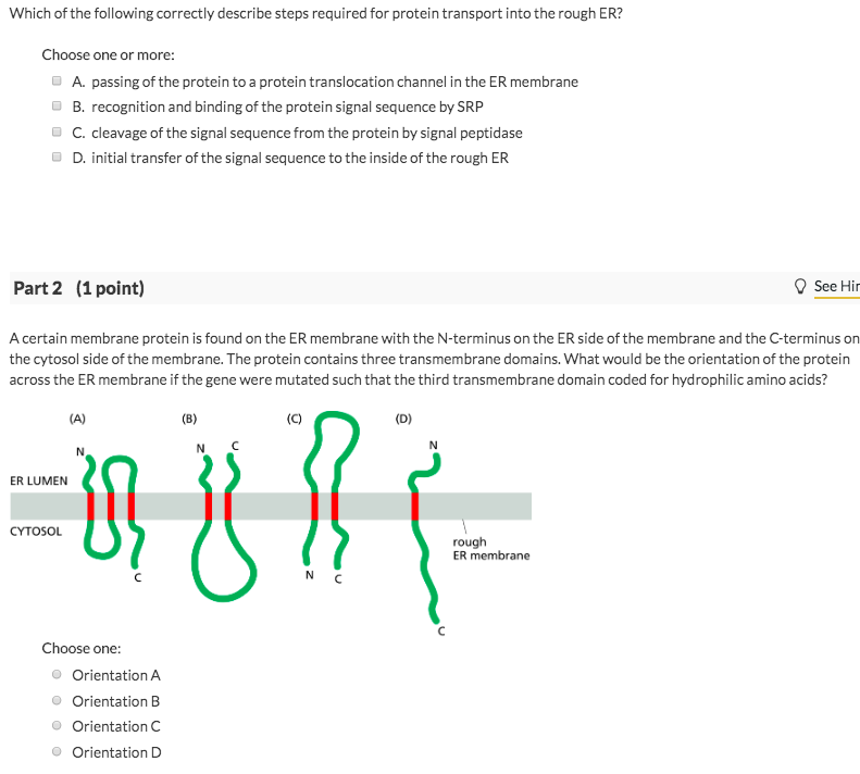
A certain membrane protein is found on the ER membrane with the N-terminus on the ER side of the membrane and the C-terminus on the cytosol side of the membrane. The protein contains three transmembrane domains. Combination of the orientation and membrane insertion provides a unique configuration of the protein-membrane complex and hence elucidates certain details of the enzyme function such as the modes of acquisition of a membrane-residing substrate and product release.
These results describe a novel workflow to define the EV proteome and the orientation of each protein including membrane protein topology. Membrane and can be applied to virtually any peripheral membrane protein or membrane-like structure. INTRODUCTION A large number of soluble proteins are transiently recruited to biological membranes during various cellular processes includingcellsignalingandmembranetraffickingTheyform.
The topology of an IMP refers to the orientation of the protein in the membrane. A protein can be in either the type I or type II topologies. Type I indicates that the N-terminus of the protein passes through the membrane first.
Integral membrane proteins function when incorporated into a lipid bilayer and they are held tightly to the lipid bilayer with the help of an annular lipid shell. Because bilayers define the boundaries of the cell and its compartments these membrane proteins are involved in many intra- and inter-cellular signaling processes. Certain kinds of membrane proteins are involved in the process of.
Protein deeply embedded in the bilayer are called integral membrane proteins. Most of this integral membrane proteins span the whole bilayer and they are called Transmembrane protein. Because many molecules and ions can not pass through the hydrophobic core of the cell membrane they need a carrier mediated transport to go in and out of the cell.
In most cases the orientation of protein in a liposomal membrane is random 11. In others is it either exclusively inside-out or outside-out relative to the native orientation 12 and cannot be tailored to a specific application.