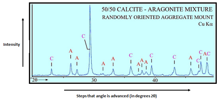
The figure below shows an example pattern of ours. A detailed explanation of how to calculate the Full Width At Half MaximumFWHM for the peaks in the XRD patternThe video demonstrates the use of Origin to.

In my opinion Search-Match the best for XRD peak identification because it simple clear easy.
How to read xrd peaks. That peak is situated at 88 and you have an indication of only illite there in blue. You have however especially marked the peak at about 125 that is suggested to contain intensity. Predicting the heights of diffraction peaks is a little more complicated and involves calculating the multiplicity of each peak the number of sets of planes related by the crystallographic symmetry as well as the structure factor which is dependent on both structure and atomic species.
A broader peak means that there may be a smaller crystal defect in the crystalline structure or that the sample might be amorphous in nature a solid lacking perfect crystallinity. For smaller samples the patterns determined using XRD analysis can be used to determine a samples composition. There is a large database of elements compounds and minerals that contain the diffraction patterns.
Results are commonly presented as peak positions at 2θ and X-ray counts intensity in the form of a table or an x-y plot shown above. Intensity I is either reported as peak height intensity that intensity above background or as integrated intensity the area under the peak. The relative intensity is recorded as the ratio of the peak intensity to that of the most intense peak relative intensity II1 x 100.
Intensity I is either reported as peak height intensity that intensity above background or as integrated intensity the area under the peak. The relative intensity is recorded as the ratio of the peak intensity to that of the most intense peak relative intensity II 1 x 100. Determination of an Unknown.
Find the Miller indices of a peak in a x-ray diffraction pattern for copper. Made by faculty at the University of Colorado Boulder Department of Chemical and. To check the nature of the materials using XRD patterns you have to look the nature of Braggs peaks appearing in the XRD pattern.
If you get a very broad humped peak then the material will be. Its better to use the software of used XRD device to identify the peaks. Also if u wish to determine the grain size u can use Williamson-Hall approach.
Also if u wish to determine the grain. The bare RGO shows diffraction peaks at 250 and 430 corresponding to 002 and 100 planes of graphite-like structure where as GO shows the diffraction peak at 100. Structure Peak identification indexing using JCPDS PCPDFWIN or COD databases - YouTube.
Maisie Williams x HM. Peak Position d-spacings and lattice parameters l 2d hklsinq hkl Fix l Cu ka 154Å d hkl 154Å2sinq hkl For a simple cubic a b c a 0 a 0 d hkl h2k2l2½ eg for NaCl 2q 22046o q 22023o d 220 19707Å a 055739Å Most accurate d-spacings are those calculated from high-angle peaks 2 2 2 0 h k l a d hkl. In my opinion Search-Match the best for XRD peak identification because it simple clear easy.
You can use filter of materials and look on information of phase hkl angular position d type. Peak can vary due to instrumental and experimental parameters. The relative intensities of the diffraction peaks should be instrument independent.
To calculate relative intensity divide the absolute intensity of every peak by the absolute intensity of the most intense peak and then convert to a percentage. The most intense peak of a phase is therefore always called the. About Press Copyright Contact us Creators Advertise Developers Terms Privacy Policy Safety How YouTube works Test new features Press Copyright Contact us Creators.
X-ray diffraction XRD is a versatile and nondestructive analytical technique that can quickly obtain detailed structural and phase information of materials. XRD analysis provides qualitative and quantitative mineral or phase information in a wide range of geological materials explored in a research laboratory or processed for an industrial application. One of the techniques our experimentalists use regularly is x-ray diffraction XRD.
XRD is a technique where you point an x-ray beam at a material in a set angle and observe the resulting angles and intensities of the diffracted beam. These patterns when measures with a camera along all angles theta result in peaks. The figure below shows an example pattern of ours.
σσσ2 GU tan2θθθθ GV tan θθθθ GW GPcos2 θθθθ sample shift. S -ππππR shft 3600 sample absorption. µµµµeff -9000 ππππR Asym peak breadth Lorentzian.
γγγγ LX -ptec cos φ φ φcos θ LY -stec cos φ φ φ φ tan θθθ Gaussian Sherrer broadening Lorentzian Sherrer broadening. A detailed explanation of how to calculate the Full Width At Half MaximumFWHM for the peaks in the XRD patternThe video demonstrates the use of Origin to.