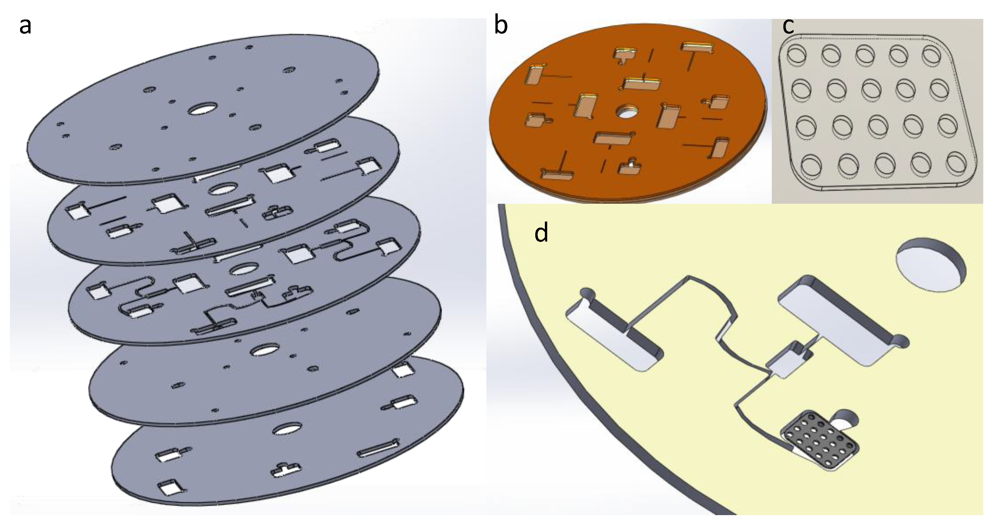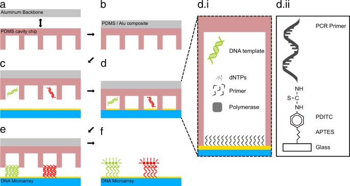
The method of spotting microarrays is achieved traditionally using a computer-controlled xyz motion stage with a head carrying a pen device to pick up small drops of solution from the multiwell plates for transfer and spotting them onto a surface. In microfluidic microarray assays the reagents are not stationary and flow.

CDNA oligonucleotides RNA peptides proteins and antibodies carbohydrates and as specialty even tissue bacteria and cells.
Microarray spotting process flow method. DNA Microarray Procedure 1 Collect Samples. This can be from a variety of organisms. Cancerous human skin tissue healthy human skin tissue.
Extract the RNA from the samples. Using either a column or a solvent such as phenol-chloroform. After isolating the RNA we need to isolate the mRNA from the rRNA and tRNA.
MRNA has a poly-A tail so we can use a column. The method of spotting microarrays is achieved traditionally using a computer-controlled xyz motion stage with a head carrying a pen device to pick up small drops of solution from the multiwell plates for transfer and spotting them onto a surface. These spotting pens are sophisticated designs adapted from the quill type of ink pen.
The pen printing is reliable and repeatable when using a flat solid. Microarrays also known as chips or biochips can be produced using one of two methods. In one method probes are synthesized separately followed by spotting onto the microarrays into very small grids by a robot.
The other method consists of synthesizing probes directly on the microarray using UV-masks and photo-activated chemistry. Microarray printers or spotters are available for. The M2-Automation systems for spotting of microarrays using Spot-on-the-Fly SotF is able to produce microarrays with a frequency of 3200 spots per second.
Spotting takes place during nozzel dispenser movement without stopping over the spot target location. A method whereby controlled arrays of hexagonally ordered monolayer islands of polystyrene particles can be deposited using a microspotter was demonstrated. The microparticle size could be varied.
Expression Microarrays The Array Thousands to hundreds of thousands of spots per square inch Each holds millions of copies of a DNA sequence from one gene Its Use Take mRNA from cells put it on array See where it sticks mRNA from gene x should stick to spot x. Microarray analysis is a technology developed in the 1990ies. It is used in interaction studies and for analysis of complex mixtures which cannot be analysed by other techniques.
The method is versatile and capable of covering the full spectrum of applications including DNA eg. CDNA oligonucleotides RNA peptides proteins and antibodies carbohydrates and as specialty even tissue bacteria and cells. Inkjet printing and microspotting methods have been used to fabricate custom microarrays by directly spotting biomolecules of interest on microscope glass slides membranes or other surfaces.
Various types of spotters and derivatized substrate surfaces have been developed to. Our microarray spotters provide the precision you need for diagnostic applications like lateral flow MTPs micro-electronics biosensors modified glass-slide surfaces cell spotting assays MALDI-MS LC-MS and Lab on a chip. The product portfolio encompasses contact and non-contact liquid handling solutions.
M2-Automations low volume pico- and nanolitre. Innovative method used to identify the associations between genetic variations and normalabnormal phenotypes especially in clinical and basic research laboratories. Allele frequencies and risk nature of the alleles differ from population to population and genotyping by microarray proves a cost-effective method to determine such.
The spotting process was carried out at 20 C and 55 humidity. After spotting the chips were incubated at 20 C and 55 humidity overnight. For microarray chip assembly the spotted chips were connected to a PMMA carrier containing in- and outlet holes using double-sided adhesive foil with cut-outs forming two flow channels.
The assembled chips were then filled with blocking. US7314595B2 - High throughput microarray spotting system and method - Google Patents High throughput microarray spotting system and method Download PDF. A protein microarray or protein chip is a high-throughput method used to track the interactions and activities of proteins and to determine their function and determining function on a large scale.
Its main advantage lies in the fact that large numbers of proteins can be tracked in parallel. The chip consists of a support surface such as a glass slide nitrocellulose membrane bead or. A common approach for performing microfluidic microarray tests is by bonding the glass microarray to the microfluidic chip.
The microarray can be aligned with microchannels in a microfluidic device such that after the bonding running the sample through the microchannels would expose the genes to the DNA spots. Large surface to volume ratio of microchannels increases the chance of pairing genes with the DNA probes. In microfluidic microarray assays the reagents are not stationary and flow.
In spotted microarrays the probes are oligonucleotides cDNA or small fragments of PCR products that correspond to mRNAs. The probes are synthesized prior to deposition on the array surface and are then spotted onto glass. A common approach utilizes an array of fine pins or needles controlled by a robotic arm that is dipped into wells containing DNA probes and then depositing each probe at designated.
Methods to generate protein microarrays by cell-free expression highlights differ-ences and similarities and reports the current state of the different approaches. Cell-free Expression DAPA NAPPA Protein microarray generation PING Additional supporting information may be found in the online version of this article at the publishers web-site Received. January 23 2014.
There are several methods for developing microarrays that include printingspotting in situ synthesizing on solid supports high-density and suspension bead arrays and electron microarrays are few of the examples. The nature and types of recognition elements are used to categorize the type of microarray. The microarray surface is coated with SHA on a 3D polymer.
The proteins or peptides are modified with PDBA and then the crude proteinPDBA complex is spotted on SHA-coated slides. The microarray technology has attained maturity and it is now possible to fabricate very high density arrays. The sensitivity has also improved and it is therefore possible to detect poorly expressed genes.