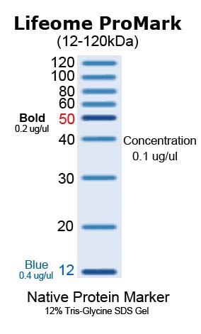
BSA is labeled with blue dye which helps in tracking purpose during electrophoresis. BSA is labeled with blue dye which helps in tracking purpose during electrophoresis.

SERVA Unstained Protein Standard 65 - 97 kDa BlueVertical PRiME Blot Modul for SERVAs BV-104 electrophoresis tank IPG tray with electrode lid for 1st dimension protein separation on 7 cm to 24 cm IPG strips Special Pharma Edition of SERVA BlueStain automated gel staining device.
Protein marker for native gel. Annons Sign Up Today For News Announcements. IEF and 2D protein standards are a mixture of native proteins with isoelectric points pI ranging from 445 to 96 providing reproducible pI calibration in native PAGE or agarose IEF gels. 2-D SDS-PAGE protein standards provide calibrated references for protein pI and molecular weight in the second dimension.
NativePAGE Bis-Tris Gels use Coomassie G-250 to bind to proteins and confers a net negative charge while maintaining the proteins in their native state without protein denaturation. The G-250 is present in the cathode buffer to provide a continuous flow of G-250 into the gel and is added to samples containing non-ionic detergent prior to loading the samples onto the gel. During native electrophoresis proteins are separated based on their charge to mass ratios.
Recommended for use with Novex NativePAGE Bis-Tris gradient Tris-Glycine or NuPAGE Novex Tris-Acetate gels. Contains markers with a wide range of high molecular weights providing 8 protein bands in the range of 201200 kDa. This Color Markers contains 6 proteins conjugated to colored dies providing a monitor for protein migration and transfer efficiency.
These markers are suitable for use in a Tricine SDS-PAGE system. Color Marker Ultra-low Range C6210 separated on a 16 Tris-tricine gel. Invitrogen Life Technologies have predesigned molecular wt markers for the native PAGE NativeMark Unstained Protein Standard Cat LC075.
Gel used for native non-denaturing gel electrophoresis of protein samples. The NativePAGE Novex Bis-Tris Gels are used with NativePAGE Running Buffers see page 5 to produce a non-denaturing electrophoresis system operating at near neutral pH. The near neutral pH environment during.
In native PAGE electrophoresis most proteins have an acidic or slightly basic pl isoelectric point 38 and migrate towards the negative polar. If your proteins pl is larger than 89 for example you should probably reverse the anode and run the native PAGE gel. New protein standards eg.
SERVA Unstained Protein Standard 65 - 97 kDa BlueVertical PRiME Blot Modul for SERVAs BV-104 electrophoresis tank IPG tray with electrode lid for 1st dimension protein separation on 7 cm to 24 cm IPG strips Special Pharma Edition of SERVA BlueStain automated gel staining device. Gel filtration markers are used in protein chromatography and gel filtration chromatography. Gel filtration markers have been used to study glucocorticoid-induced lymphocytolysis as a model system for apoptosis within the immune system.
Gel filtration markers have also been used to isolate Polygonatum odoratumlectin which showed. Protein molecular weight markers typically used polyacrylamide gel electrophoresis and Western blotting. Rainbow markers Mr 3500 to 225 000.
Prestained visible marker of different colors in gels and Western blots. Sodium dodecyl sulfate SDS-PAGE Western blot. Originally molecular weight markers were a mixture of easy-to-purify proteins of known molecular weight.
These marker proteins were unstained and were generally visualized on SDS-PAGE gels by staining the gel with Coomassie Brilliant Blue R250 or after western transfer by a stain such as Ponceau S. Now there are a myriad of options. Native PAGE Protein molecular weight marker is a set of five proteins with molecular weight ranging from 240kD to 184kD to characterize the proteins separated in poly-acrylamide gels in their native state.
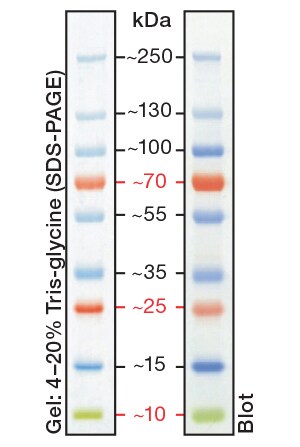
BSA is labeled with blue dye which helps in tracking purpose during electrophoresis. Protein MW Da Recommended of gel. Load 5 µl PageRuler Prestained Protein Ladder into marker narrow well Fig.
Cut the gel strip such that it easily fits into the 2D well. Mark the relevant positions corresponding to the expected protein sizes on the gel cassette. Add 100 μl of 1 protein loading buffer on top of gel.
A protein marker also called a protein molecular weight marker a protein MW marker or a protein ladder is used to estimate the size of proteins resolved by gel electrophoresis. All markers are optimized for use with LI-COR imaging systems but can be used with other imagers. LI-COR protein ladders and markers are visible on the gel during.
Several changes of Destaining Solution the gel background becomes colorless and leaves protein bands colored blue purple or red. Coomassie Brilliant Blue R250 and PAGE Blue 83 each visibly stain as little as 01-1 ug of protein. Annons Sign Up Today For News Announcements.