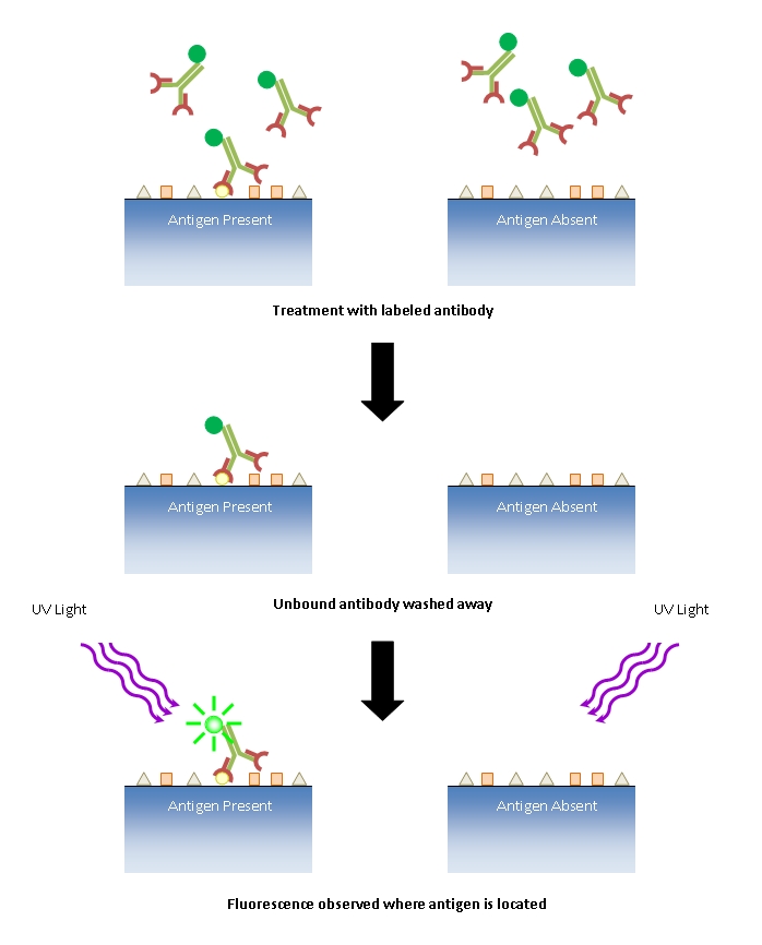
First adjust antibody concentration. Incubate cells with 1 BSA 2252 mgmL glycine in PBST PBS 01 Tween 20 for 30 min to block unspecific binding of the antibodies alternative blocking solutions are 1 gelatin or 10 serum from the species the secondary antibody was raised in typically goat serum or donkey serum.

Incubate cells with 1 BSA 2252 mgmL glycine in PBST PBS 01 Tween 20 for 30 min to block unspecific binding of the antibodies alternative blocking solutions are 1 gelatin or 10 serum from the species the secondary antibody was raised in typically goat serum or donkey serum.
Role of blocking in immunofluorescence. In this method blocking is done with normal unchallenged serum from the same species that the secondary antibody was raised in. Normal serum carries antibodies that will bind to the non-specific epitopes in your sample thus blocking your conjugated antibodies from doing the same. Blocking of the direct immunofluorescence IF reaction al-lowed titration of anti-membrane antigen MA or anti-intracellular antigen IA antibodies induced by herpesvirus of turkeys HVT.
The titers blocking index obtained by the blocking IF test were well correlated with those determined by. Blocking Strategies for IHC. Before using specific antibodies to detect antigens by immunohistochemistry IHC all potential nonspecific binding sites in the tissue sample must be blocked to prevent nonspecific antibody binding.
If blocking is omitted or inadequate the antibodies or other detection reagents may bind to a variety of sites that. Incubate cells with 1 BSA 2252 mgmL glycine in PBST PBS 01 Tween 20 for 30 min to block unspecific binding of the antibodies alternative blocking solutions are 1 gelatin or 10 serum from the species the secondary antibody was raised in typically goat serum or donkey serum. See antibody datasheet for recommendations.
Controls to convince you of the specificity are important eg secondary only blocking primary antibody with the proteinpeptide against which it is raised lack of signal in knockoutRNAi cells. Lastly because your IF results will be of only a few cells it. Scribe the slides while the block is in the cryostat in case you happen to break one and eventually everybody does at one time or another that way the block is still faced up to the knife edge.
You can pick up new sections on another slide and keep moving forward. Cells grown on cover slips or on commercially available incubation chambers. PBS and cPBS complete PBS 2.
Fixative 4 formaldehyde in PBS freshly prepared 3. 50 mM NH4Cl in PBS or 01M Glycine in PBS 4. Blocking solution 1 BSA or 10 FCS fetal calf serum in PBS 5.
Immunohistochemistry IHC is the most common application of immunostainingIt involves the process of selectively identifying antigens proteins in cells of a tissue section by exploiting the principle of antibodies binding specifically to antigens in biological tissues. IHC takes its name from the roots immuno in reference to antibodies used in the procedure and histo meaning tissue. The western blot is extensively used in biochemistry for the qualitative detection of single proteins and protein-modifications such as post-translational modificationsAt least 8-9 of all protein-related publications are estimated to apply western blots.
It is used as a general method to identify the presence of a specific single protein within a complex mixture of proteins. The reason we use this for all our incubations is because of our secondary antibodies. We use donkey secondary antibodies and thus any source of non-specific staining or background will be best blocked with donkey serum.
That way anything your antibodies will bind in. Whole-mount immunofluorescence staining is intended for smaller sections of tissue without the need for manual sectioning. To this purpose the mouse heart is dissected with unwanted tissue removed followed by fixation permeabilization and blocking.
Cells of the conduction system within SAN and AVN are then stained with an anti-HCN4 antibody. HEQDBASRHU Role of Immunofluorescence in Skin Lesions Kindle You May Also Like PDF Crochet. Learn How to Make Money with Crochet and Create 10 Most Popular Crochet Patterns for Sale.
Learn to Read Crochet Patterns Charts and Graphs Beginner s Crochet Guide with Pictures. To reduce non-specific antibody binding membranes were incubated for 2 h at room temperature in bovine serum albumin BSA-based blocking buffer. The membranes were then exposed to primary antibodies overnight at 4C.
Only those antibodies recommended for WB were tested. Protein blocking for IHC. Blocking with sera or a protein blocking reagent prevents non-specific binding of antibodies to tissue or to Fc receptors.
Theoretically any protein that does not bind to the target antigen can be used for blocking. In practice some proteins bind more readily to non-specific sites. Serum is a common blocking agent as.
METHOD SUMMARY A brief slow-freezing step is added to immunofluorescence procedures in order to improve antibody penetration while maintaining physiological sample structure. Brief freezing steps lead to robust immunofluorescence in the Drosophila nervous system. Detection of human adenoviruses HAdVs in nasopharyngeal swab samples by immunofluorescence assay IFA will be valuable for diagnosing HAdV infection which is a leading cause of severe respiratory tract disease and will help in curbing the spread of HAdV.
DNA 5-hydroxymethylcytosine 5hmC is an important epigenetic modification found in various mammalian cells. Immunofluorescence imaging analysis essentially provides visual pictures for the abundance and distribution of DNA 5hmC in single cells. However nuclear DNA is usually wrapped around nucleosomes packaged into chromatins and further bound with many functional proteins.
If there is the background in the results of the immune fluorescence we should first consider whether the primary antibody concentration or the concentration of the second antibody is too high. First adjust antibody concentration.