Phase Separation in Lipid Lamellae Result from Ceramide Conformations and Lateral Packing Structure Hiroki Ohnari a Eiji Naru Taku Ogurab Osamu Sakataa and Yasuko Obatac. The second stage of tearing is the formation of crack terraces by the linking together of individual separations.

The lamellarnon-lamellar transformations play an important role in.
Stage of lamellar separation. Lamellar separation is now shown by electron microscopy to be due to folds crossing the lens fibres. The clinical study showed the lines occurring with spoke cataract and the electron microscopy showed the association with the novel finding of peripheral breaks in. Lipid concentration is applied and the lamellar phase separation to two or three phases is induced in the vicinity of the critical unbinding point 13.
In the theory salt concentration possibly become the second-order parameter and could be the origin of the lamellar phase separation. A lamellar tear takes place in three stages as. Start of separation and void formation at inclusions in the base metal.
Formation of crack terraces by the linking together of individual separations. Joining of terraces through shear failure. The tear itself exhibits a characteristic terraced profile and it is this step-like terrace look that distinguishes a lamellar tear from an.
Lamellar separation is seen as parallel lines in the lens cortex. It has been the subject of a joint study between Oxford and Amsterdam. The condition was studied in vivo by macro photography and.
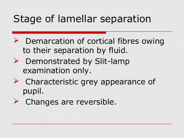
Stage of lamellar separation Demarcation of cortical fibres owing to their separation by fluid Demonstrated by Slit-lamp examination only Characteristic grey appearance of pupil Changes are reversible. Therefore freeze-induced lamellar-to-hexagonalII phase transitions in the plasma membrane are a consequence of dehydration rather than subzero temperature per se. When suspensions of protoplasts isolated from cold-acclimated leaves were frozen to – 10 degrees C no injury was incurred and hexagonalII phase transitions were not observed.
The unique anatomy of the fovea may make it more likely to develop an outer layer separation from traction on the inner retina Figure 15-5 which can progress to a separation of the photoreceptor layer to form an outer lamellar hole Figure 15-6 and finally dehiscence of the inner retina to produce a full-thickness hole Figure 15-7. Stage of Lamellar separation Demarcation separation of Cortical fibers by Fluid. Stage of Incipeint Catract Cuneiform.
Wedge shaped Opacities Extend from peripheral to central Radial spoke like pattern on Oblique illumination Dark lines against Red fundus on DDO Late loss of Visual acuity. Lamellar hexagonal cubic etc struc-tures rather than the lamellar state. Consequently except for the lamellar liquid-crystalline phase the mem-brane phase the membrane lipids are also able to form a large variety of other phases with different geom-etry and molecular structure.
Lamellar bodies at various stages of development in type II cells of fetal mouse lung. Well-developed multivesicular bodies and particulate glycogen arrows are characteristic of this stage of type II cell differentiation. Figure 2 Fusion of a multivesicular body with a lamellar body in a fetal mouse type II cell.
Lamellar sedimentation is based on the principle that in free settling according to Hazens law see different types of sedimentation granular particle retention is independent of the structures heightTherefore the surface area available for sedimentation can be extended quite considerably by superimposing a large number of watersludge separation cells on top of the. A lamella clarifier or inclined plate settler IPS is a type of settler designed to remove Particulates from liquids. They are often employed in primary water treatment in place of conventional settling tanksThey are used in Industrial water treatmentUnlike conventional clarifiers they use a series of inclined plates.
These inclined plates provide a large effective settling area for a. Failure results in delamination separation of lamellae and may be one of the initial stages of herniation and degeneration68 Knowledge of the ILM structure and mechanical function is important to determining the loading conditions under which the AF is at risk of delamination and subsequent disc disruption and herniation. The term lamellar hole was used to describe both the end stage of a cystoid macular edema 14 and the aborted process of formation of a macular hole.
44 66 With the advent of high resolution or spectral domain OCT the thinning of the foveal center due to the contraction of an epiretinal membrane was also referred to as a lamellar macular. In protoplasts isolated from nonacclimated rye leaves Secale cereale L. Cultivar Puma cooling to – 10 degrees C at a rate of 1 degrees Cmin results in extensive freeze-induced dehydration osmotic contraction and injury is manifested as the loss of osmotic responsiveness during warming.
Under these conditions several changes were observed in the freeze-fracture morphology of the plasma. First was a stage of early lamellar separation where small intralenticular clefts were noted superficially. Second was the stage of established lamellar separation where crescentic fluid clefts appeared interspersed between the lens fibers and the depth increased as a function of severity.
Dispersions of double-chain non-lamellar membrane lipids frequently display a lamellarinverted hexagonal L α H II phase transition. In some instances they can also form inverted phases of cubic symmetry. The lamellarnon-lamellar transformations play an important role in.
A lamellar tear takes place in three stages. In initiates by separation and void formation at inclusions in the base metal. The second stage of tearing is the formation of crack terraces by the linking together of individual separations.
Phase Separation in Lipid Lamellae Result from Ceramide Conformations and Lateral Packing Structure Hiroki Ohnari a Eiji Naru Taku Ogurab Osamu Sakataa and Yasuko Obatac. The lamellar structure of the V-shaped CER conformation has a low orthorhombic ratio. The above results suggest that an increase in the ratio of CER with the V.